Physical Therapy Toolbox: radial and ulnar arteries


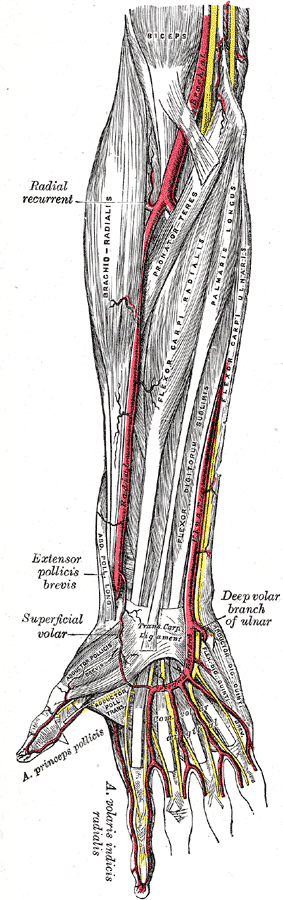
The image shows some of the muscles and arteries of the right forearm and hand, including the superficial palmar arch (titled Superficial Volar Arch in this picture, which is an alternative term) and the common palmar digital arteries branching off of it. The unlabelled yellow lines are nerves. Palmar aspect with the proximal part (elbow) at the top and the distal part (hand) at the bottom.


Copyright © 2008 Physical Therapy Working for You. Contact us    Disclaimer


Copyright (c) 2008 Physical Therapy Working For You. Permission is granted to copy, distribute and/or modify this document under the terms of the GNU Free Documentation License, Version 1.2 or any later version published by the Free Software Foundation; with no Invariant Sections, no Front-Cover Texts, and no Back-Cover Texts. A copy of the license is included in the section entitled "GNU Free Documentation License". Wikipedia

Images are a faithful reproduction of a lithograph plate from Gray's Anatomy, a two-dimensional work of art, that is not copyrightable in the U.S. as per Bridgeman Art Library v. Corel Corp. Unless stated otherwise, it is from the 20th U.S. edition of Gray's Anatomy of the Human Body, originally published in 1918 and therefore lapsed into the public domain.